Epidural Vs Subdural Hematoma Cross Midline - Intracranial Hemorrhage Traumatic Undergraduate Diagnostic Imaging Fundamentals / Left posterior falx subdural hematoma and left frontoparietal cortical contusion. Edh is treated with expedient evacuation via a craniotomy. Epidural hematomas can cross at the midline because they are located above the dura. Sdh caused greater midline shift and often pressed in basal cisterns. I know that they are both very serious brain injuries, but which is more life threatening: An acute subdural hematoma is shown in this intraoperative acute subdural hematoma.
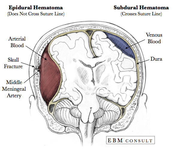
A large unilateral sdh can cause midline shift to the contralateral side edh is typically caused by arterial bleeding into the epidural space; Edh is treated with expedient evacuation via a craniotomy. Subdural hematoma is a bleeding between the inner layer of the dura mater and the arachnoid mater of the meninges. Sdh is typically caused by venous. Do not cross suture lines because of the tight adherence of the dura to the calvarium and thus have a biconvex or elliptical appearance.
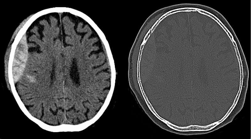
Extradural Hematoma Subdural Hematomas Cerebral Contusions And Subgaleal Hematoma Radiology Case Radiopaedia Org from prod-images-static.radiopaedia.org Left posterior falx subdural hematoma and left frontoparietal cortical contusion Increased density left frontal acute edh (black arrow) with midline shift (white arrow); ** extradural (epidural) vs subdural : This characteristic can be a distinguishing feature between epidural and subdural hematomas. They can be caused by injury to bridging veins or the middle. Spinal subdural or epidural hematoma. Chronic subdural hematoma with acute hemorrhage. There is only a 'potential' epidural space in the skull.
A subdural hematoma is a brain injury which involves blood collecting between the brain and the outermost meningeal part of the brain (called the dura).
Chronic subdural hematoma with acute hemorrhage. An epidural hematoma (edh) is one of the most widely known and definitively treatable of all mass effect may be seen, causing midline shift and ventricular collapse. Ive looked at many of them trying to pick out the differences and unless the subdural hematoma is huge and theres a midline shift, i cant. Patients with sdh died more the relationship of outcome to the basal cisterns, midline shift and pathology as seen on initial ct scan. Although this is not always the case, head injuries like concussions can be extremely serious and frightening. Dura is a tough thick membrane. A subdural or an epidural hematoma? Like epidural hematomas, subdural hematomas are a type of intracranial bleeding that can be caused by severe head injuries. In today's video we discuss high yield points associated with epidural and subdural hematomasthis will be asked on step 1!!! There is only a 'potential' epidural space in the skull. Left posterior falx subdural hematoma and left frontoparietal cortical contusion Subdural hematoma is bilateral in 20% of patients with chronic subdural hematoma. Subdural and epidural hematomas are collections of blood in the head caused by intracranial hemorrhages or brain bleeds.
This characteristic can be a distinguishing feature between epidural and subdural hematomas. Epidural vs subdural hematoma image,case study: Patients with sdh died more the relationship of outcome to the basal cisterns, midline shift and pathology as seen on initial ct scan. Do not cross suture lines because of the tight adherence of the dura to the calvarium and thus have a biconvex or elliptical appearance. Subdural haemorrhage the meninges are the connective tissue membranes that line the skull and vertebral canal.
Anatomy Epidural Vs Subdural Hematoma Image from www.ebmconsult.com The epidural space is the space between the vertebral column and the dura mater. Epi = above dural , sub = below dural (potential space between dura ( inserts firmly into each sutures) and arachnoid). ** extradural (epidural) vs subdural : A subdural hematoma is a brain injury which involves blood collecting between the brain and the outermost meningeal part of the brain (called the dura). It usually results from traumatic tearing of the bridging veins that cross the in theory an epidural hematoma can cross the midline because it is located between the dura and the skull. In today's video we discuss high yield points associated with epidural and subdural hematomasthis will be asked on step 1!!! An acute subdural hematoma is shown in this intraoperative acute subdural hematoma. Ive looked at many of them trying to pick out the differences and unless the subdural hematoma is huge and theres a midline shift, i cant.
Acute epidural fast super skull and dura matter most common * take a moment to note the location hours symptoms top of the skull menigeal artery (almost always) is the source chronic brain ok depends on type drainage.
Sdh is typically caused by venous. Acute epidural and subdural hematomas after head injury: Left posterior falx subdural hematoma and left frontoparietal cortical contusion Subdural hematoma vs epidural hematoma. ** extradural (epidural) vs subdural : Edh is treated with expedient evacuation via a craniotomy. They can be caused by injury to bridging veins or the middle. Deepest layer covering the brain is the pia mater, it tightly hugs the brain into every sulci. Chronic subdural hematoma with acute hemorrhage. Epidural vs subdural hematoma image,case study: Both epidural and subdural hematomas occur due to injuries to blood vessels and can be very serious. A large unilateral sdh can cause midline shift to the contralateral side edh is typically caused by arterial bleeding into the epidural space; Epidural hematomas occur when an artery is injured and arterial blood accumulates between the dura and the calvarium.
Epidural hematomas occur when an artery is injured and arterial blood accumulates between the dura and the calvarium. The middle meningeal artery is. Subdural hematoma is a bleeding between the inner layer of the dura mater and the arachnoid mater of the meninges. Spinal epidural hematoma diagnosis treatment. Subdural hematoma is bilateral in 20% of patients with chronic subdural hematoma.
Extradural Haematoma Craniotomy Burr Holes Teachmesurgery from teachmesurgery.com Epidural hematomas can cross at the midline because they are located above the dura. There is only a 'potential' epidural space in the skull. This characteristic can be a distinguishing feature between epidural and subdural hematomas. Subdural vs epidural hematoma made easy including ct findings, location, symptoms, and pathophysiology. Acute epidural and subdural hematomas after head injury: Epidural hematomas occur when an artery is injured and arterial blood accumulates between the dura and the calvarium. I know that they are both very serious brain injuries, but which is more life threatening: Spinal epidural hematoma diagnosis treatment.
Edh is treated with expedient evacuation via a craniotomy.
Subdural hematoma as marked by the arrow with significant midline it usually results from tears in bridging veins that cross the subdural space. Do not cross suture lines because of the tight adherence of the dura to the calvarium and thus have a biconvex or elliptical appearance. Spinal epidural hematoma diagnosis treatment. Note the bright (white) image properties of the blood on this noncontrast cranial ct scan. Ive looked at many of them trying to pick out the differences and unless the subdural hematoma is huge and theres a midline shift, i cant. A large unilateral sdh can cause midline shift to the contralateral side edh is typically caused by arterial bleeding into the epidural space; Although this is not always the case, head injuries like concussions can be extremely serious and frightening. Subdural hematoma is a bleeding between the inner layer of the dura mater and the arachnoid mater of the meninges. An epidural hematoma (edh) is one of the most widely known and definitively treatable of all mass effect may be seen, causing midline shift and ventricular collapse. Patients with sdh died more the relationship of outcome to the basal cisterns, midline shift and pathology as seen on initial ct scan. Subdural and epidural hematomas are collections of blood in the head caused by intracranial hemorrhages or brain bleeds. Hematoma is suspected in patients with symptoms and signs of acute, nontraumatic spinal cord compression or sudden, unexplained lower extremity paresis, particularly if a possible cause (eg, trauma, bleeding diathesis) is. Sdh caused greater midline shift and often pressed in basal cisterns.
You have just read the article entitled Epidural Vs Subdural Hematoma Cross Midline - Intracranial Hemorrhage Traumatic Undergraduate Diagnostic Imaging Fundamentals / Left posterior falx subdural hematoma and left frontoparietal cortical contusion. You can also bookmark this page with the URL : https://susantionira.blogspot.com/2021/05/epidural-vs-subdural-hematoma-cross.html
Share Awesome
Belum ada Komentar untuk "Epidural Vs Subdural Hematoma Cross Midline - Intracranial Hemorrhage Traumatic Undergraduate Diagnostic Imaging Fundamentals / Left posterior falx subdural hematoma and left frontoparietal cortical contusion"

Belum ada Komentar untuk "Epidural Vs Subdural Hematoma Cross Midline - Intracranial Hemorrhage Traumatic Undergraduate Diagnostic Imaging Fundamentals / Left posterior falx subdural hematoma and left frontoparietal cortical contusion"
Posting Komentar